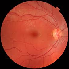
At Back of the Eye M.D., Dr. Saralyn Notaro Rietz specializes in an array of areas. We also offer many services, one of these being fundus photography. So what exactly is the fundus? The fundus refers to the innermost surface of the eye, and includes the retina, the optic nerve, and blood vessels. Match that with photography, and you have a photographic document of the retina, also known as the back portion of the eye.
Not only is photography used to document findings, but also to compare conditions from visit to visit. Fundus photos can be key to detecting and following several diseases such as Macular Degeneration, Diabetic Retinopathy, and Retinal Detachments, to name a few.
Our office also offers autofluorescence photography, which is a unique type of photograph. Fundus autofluorescence photography is a way to help the doctor attain a better understanding of the processes going on beneath the retina. The benefits? Obtaining earlier diagnoses and the ability to predict the movement of some retinal diseases. It is particularly useful in Macular Degeneration.


